Breast Tomosynthesis (3D Mammography)
The Jackson Clinic now offers Breast Tomosynthesis or 3D Mammography for breast cancer screening. Breast Tomosynthesis mammography produces a three-dimensional view of the breast tissue that helps radiologists identify and characterize individual breast structures without the confusion of overlapping tissue.

The benefits of Breast Tomosynthesis include:
- 41% increase in the detection of invasive breast cancers
- 29% increase in the detection of all breast cancers
- 15% decrease in women recalled for additional imaging
- 49% increase in Positive Predictive Value (PPV) for a recall
- 21% increase in PPV for biopsy.
The Breast Tomosynthesis or 3D Mammography will benefit all screening and diagnostic mammography patients and is especially valuable for women receiving a baseline screening, those who have dense breast tissue and women with a personal history of breast cancer.
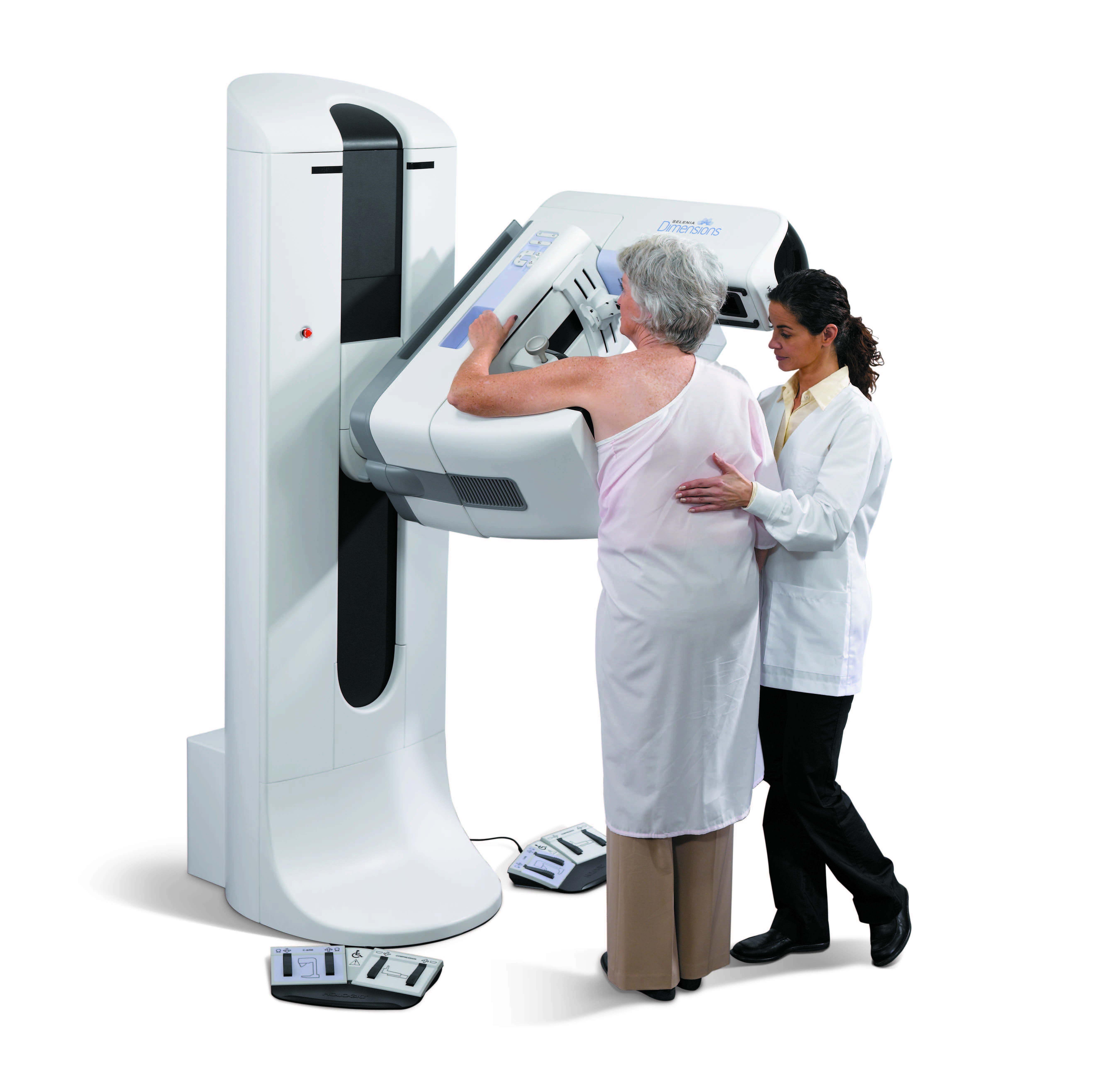
A Breast Tomosynthesis screening experience is similar to a traditional mammogram. During the mammography exam, multiple, low-dose images of the breast are acquired at different angles. These images are then used to produce a series of one-millimeter thick slices that can be viewed as a 3D reconstruction of the breast.
Frequently Asked Questions
Now that 3D mammography is available at our facility, you may have some questions. We’ve prepared this short Q&A to address concerns you may have.
What is a 3D mammography breast exam?
3D mammography is a revolutionary new screening and diagnostic tool designed for early breast cancer detection that can be done in conjunction with a traditional 2D digital mammogram. During the 3D part of the exam, the X-ray arm sweeps in a slight arc over your breast, taking multiple breast images. Then, a computer produces a 3D image of your breast tissue in one millimeter slices, providing greater visibility for the radiologist to see breast detail in a way never before possible. They can scroll through images of your entire breast like pages of a book. The additional 3D images make it possible for a radiologist to gain a better understanding of your breast tissue during screening1 and the confidence to reduce the need for follow-up imaging.
Why is there a need for tomosynthesis breast exams? What are the benefits?
With conventional digital mammography, the radiologist is viewing all the complexities of your breast tissue in a one flat image. Sometimes breast tissue can overlap, giving the illusion of normal breast tissue looking like an abnormal area. By looking at the breast tissue in one millimeter slices, the radiologist can provide a more confident assessment.1 In this way, 3D mammography finds cancers missed with conventional 2D mammography. It also means there is less chance your doctor will call you back later for a second look because now they can see breast tissue more clearly.
What is the difference between a screening and diagnostic mammogram?
A screening mammogram is your annual mammogram that is done every year. Sometimes the radiologist may ask you to come back for follow-up images which is called a diagnostic mammogram to rule out an unclear area in the breast or if there is a breast complaint that needs to be evaluated.
What should I expect during the 3D mammography exam?
3D mammography complements standard 2D mammography and is performed at the same time with the same system. There is no additional compression required, and it only takes a few more seconds longer for each view.
Is there more radiation dose?
Very low X-ray energy is used during the exam, just about the same amount as a traditional mammogram done on film.
Who can have a 3D mammography exam?
It is approved for all women who would be undergoing a standard mammogram, in both the screening and diagnostic settings.
Please call 731-422-0213 to schedule your annual screening mammogram appointment or visit www.jacksonclinic.com


